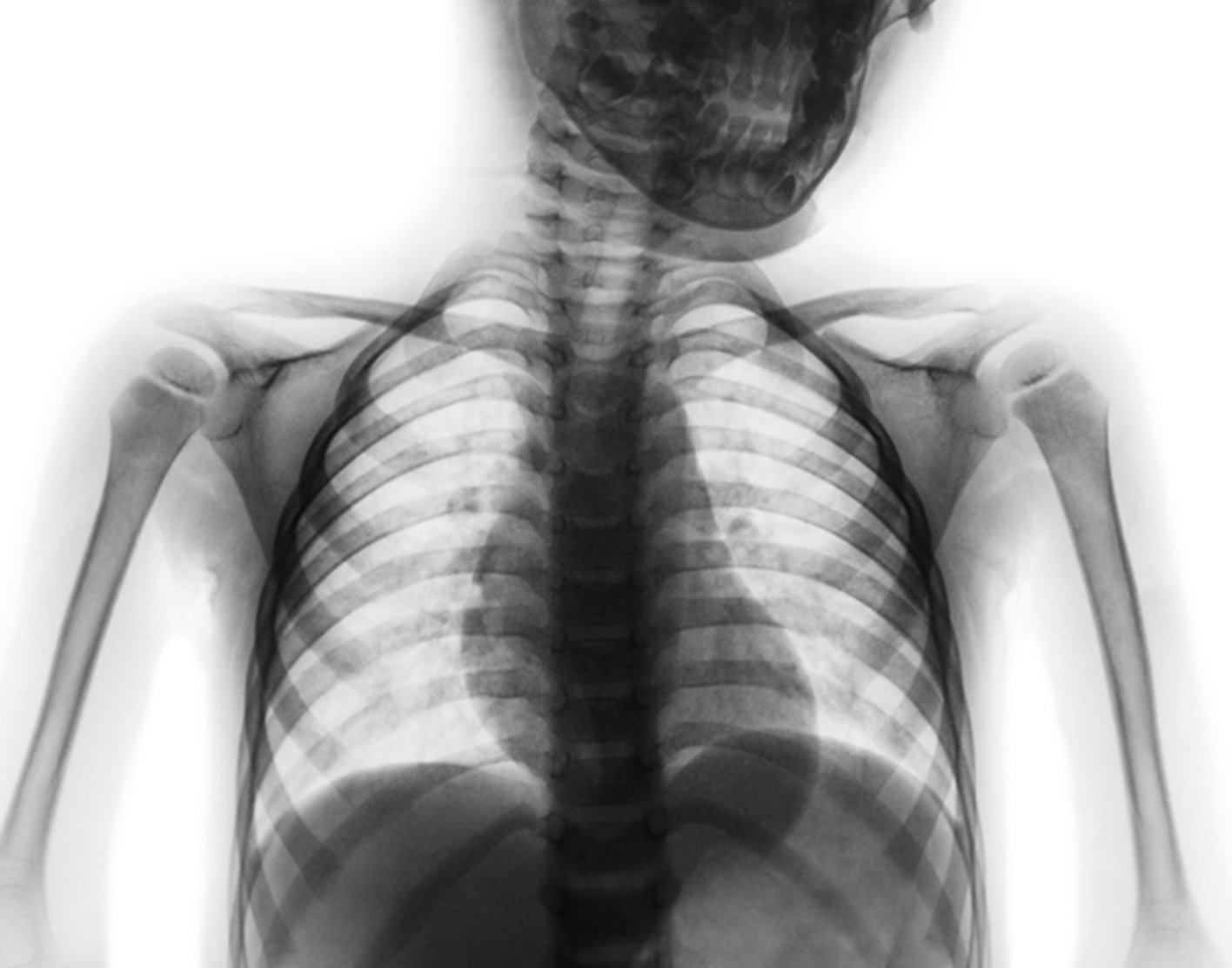
Thoracic spine x-ray
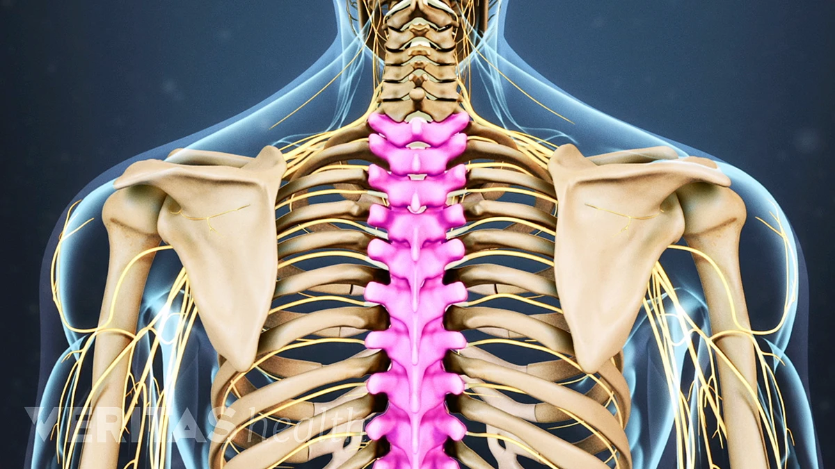
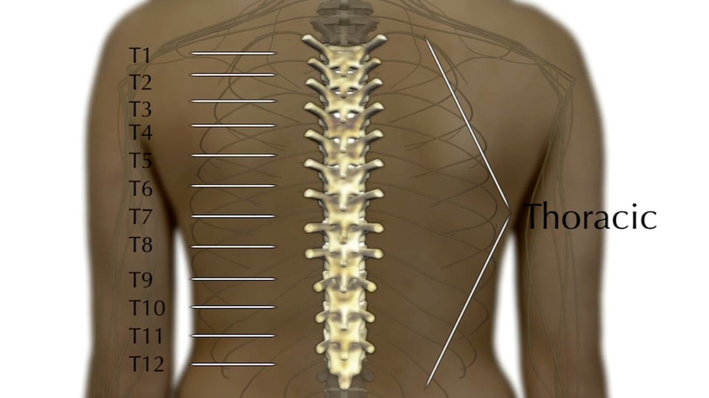
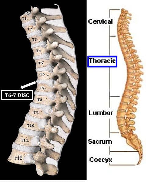
A thoracic spine x-ray is an x-ray of the twelve chest (thoracic) bones (vertebrae). The vertebrae are separated by flat pads of cartilage called disks.

Initial plain X-rays of the thoracic spine showing that the dislocated

Osteoarthritis of the thoracic spine C017 / 0697 For sale as Framed Prints, Photos, Wall Art and Photo Gifts

Thoracic Spine X-ray - W-Radiology

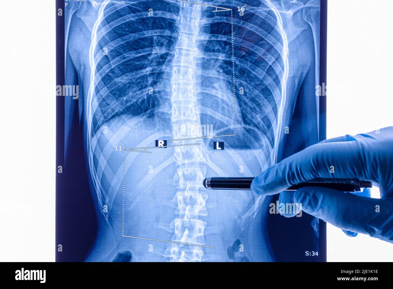
File:Thoracic spine AP.png - Wikipedia
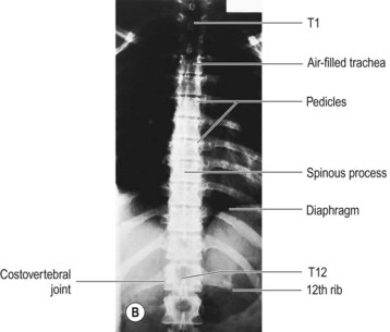
Thoracic spine

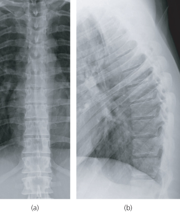
Thoracic spine X-ray examination of patients with back pain using different breathing technique and exposure times – A diagnostic study - ScienceDirect

Normal pediatric thoracic spine series, Radiology Case
Thoracic and Lumbar Spine
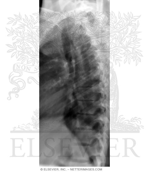
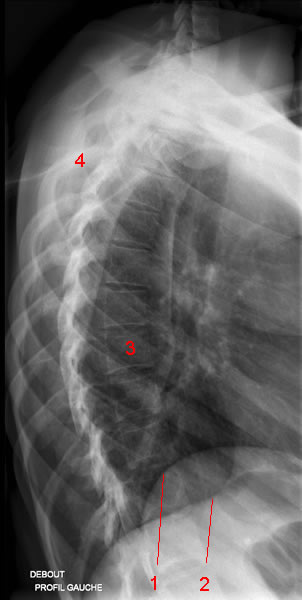
Lateral X-Ray of the Thoracic Spine

Spinal x-ray (a) and MRI (b) of the thoracic spine. Fig. 1a: The spinal

Thoracic spine fracture, X-ray and CT scan - Stock Image - C052/9109 - Science Photo Library
Thoracic Spine X-ray - W-Radiology

Frontal X-ray Thoracic Spinal Instrumentation - Stock Image - C043/0334 - Science Photo Library
Spine xray doctor hi-res stock photography and images - Alamy

The Thoracic Spine MRI Report: What You Need to Know, by Ponea